
Chest CT Image Analysis
Chest CT related outcome measures are becoming increasingly important for both clinical research and trials in cystic fibrosis (CF). Bronchiectasis, mucus plugging, and trapped air have been well-validated as outcome measures in CF, but traditional scoring systems are time-consuming and semi-quantitative. LungAnalysis co-developed and validated a sensitive alternative image analysis method, PRAGMA-CF, to score chest CT in both children and adults. PRAGMA-CF has now been successfully used as a primary outcome measure in two large studies (RCTs in young children) and has proven to be significantly more sensitive in detecting early CF lung changes compared to previously used semi-quantitative scoring methods.
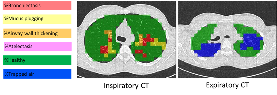
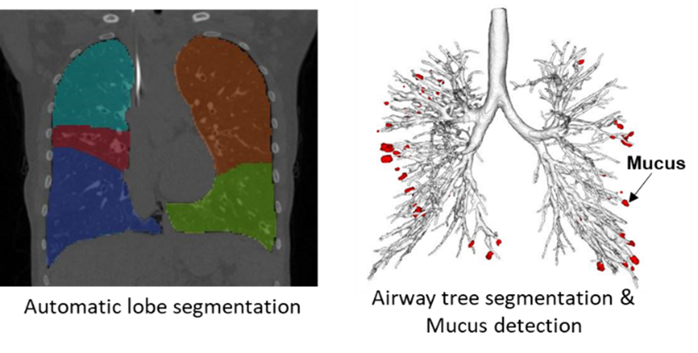
LungAnalysis has been working towards developing automated tools to quantify lung structural abnormalities. The lab has ongoing collaborations with Thirona B.V. (Nijmegen), a Dutch company specializing in cutting-edge artificial intelligence strategies for quantitative lung imaging. Together, LungAnalysis and Thirona co-developed a fully automated, sensitive system to measure bronchial widening, bronchial thickening, mucus plugging, and trapped air on chest CT. The algorithm has been integrated into Thirona’s certified software platform, LungQTM, which is installed on the virtual platform DRE at LungAnalysis, Erasmus MC. Current work on the validation of the automated tools is ongoing in a wide range of lung diseases, including CF.
Accurate imaging-based outcome measures are critical not only for clinical research and trials but also for patient registries. In 2022, LungAnalysis was awarded a large grant by the US CF Foundation (CFF) for the ENRICH project to analyze up to 10,000 CTs and incorporate chest CT related outcome measures into the ECFS Patient Registry. A similar study is currently underway for the Bronchiectasis Registry (the EMBARC study) at LungAnalysis.
Standardization of Chest CT Scanning Protocols
As chest CT involves ionizing radiation, it is essential to standardize chest CT scanning protocols to optimize the balance between radiation dose and diagnostic quality. To ensure robust and reproducible results in quantitative image analysis, chest CT images must be acquired in a standardized manner with adequate image quality.
As the chest CT core laboratory, LungAnalysis aims to standardize chest CT image acquisition across all ECFS-CTN sites and other external sites, reducing barriers to conducting clinical studies that include chest CT related outcome measures. Additionally, standardizing routine chest CT scans performed at ECFS-CTN sites for monitoring CF lung disease will facilitate the use of automated image analysis. This advancement will make it possible to integrate chest CT outcomes into the ECFS patient registry.
Standardization of Lung Volume
For the acquisition of chest CTs in cooperative subjects, for both clinical research and trials, it is crucial to standardize lung volume. Inspiratory chest CT should be acquired at a lung volume near total lung capacity (TLC), while expiratory scans should be performed at a volume near residual volume (RV). As part of the SCIFI studies, LungAnalysis assisted all ECFS-CTN sites in implementing volume-controlled chest CT for both clinical research and trials.